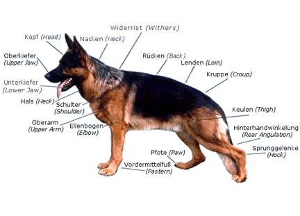
There are an average total of about three hundred and nineteen bones in the dog
skeleton. The bones have their own supply of blood vessels and nerves. They are composed of minerals, primarily calcium and
phosphorus. Bones provide framework for the body and protect internal organs. Other bones such as the bones of the legs grow
from areas of immature bone located near the ends. They are called growth plates. The growth plates provide growth to the
bone that grow and add length to the bone until the puppy’s bone is complete. This usually happens by the time that
the puppy turns the age of one. Then the plates turn hard with calcium. Immature bone growth causes bones to be weak around
the wrist (Carpus) and knee (Stifle).
Now the functions of the muscles are also important to the movement to all or a part of the
body’s body. There are two types of muscles: Smooth and Striated. Smooth muscles are found within internal organs such
as the intestines, stomach and bladder. They are muscles that function automatically. Striated muscles on the other hand are
muscles that are attached to the skeleton and they are voluntary muscles.
Muscles are connected to the bones by tough fibrous bands called tendons.
Tendons begin on a muscle and end on a bone. Ligaments connect bone to bone and span across joints. Joints are places where
two bones meet covered by a layer of smooth cartilage. A joint consists of bones, muscles, ligaments, cartilage and a lubricating
joint fluid all enclosed by a tough joint capsule.
The Digestive system:
The Digestive system includes the mouth, esophagus, stomach, intestines, liver and pancreas.
The esophagus is a small tube, which connects the mouth to the stomach. As it leaves the mouth, it follows a straight path
through the neck and chest through the diaphragm muscles and finally enters the stomach. In the dog tit takes five seconds
for food to move from the mouth to the stomach. The stomach is designed to store food and begin the digestive process. The
esophagus carries food to the stomach where it enters the cardiac sphincer. On the Interior surface of the stomach is a series
of folds called gastric folds. The folds help grind and digest foods. The inner stomach lining secretes acids and enzymes
to break food down as the initial step of the digestive process. Once the stomach digestions is complete the partially digested
food exits the stomach through the Pyloric sphincter and then enters the small intestines (duodenum) Food leaves the stomach
twelve hours after food is digested.
The small intestine is a tube like structure extending between the stomach and large intestine.
It’s the largest portion of the digestive tract and is two and a half times the animal’s total body length. An
animal that is twenty-four inches long would have an intestine sixty inches long. The small intestine has three parts. The
first part attaches to the stomach, it is known as the duodenum. In a forty-pound dog it is ten inches long. The middle (the
longest) is the jejunum. The shortest is the Ileum, connects to the large intestine. The Duodenum has important functions.
The Gall bladder and pancreas connect by the bile and pancreatic ducts. Enzymes and other secretions are important for digestion
and are produced by the liver and pancreas and pass through the ducts to mix with the food in the duodenum. The jejenum is
the longest of the small intestine and is full of villi, which absorb nutrients. Intestinal contents of the jejenum empty
into the Ileum and from there pass into the colon.
The large intestine connects to the small to the anus. The large is about sixteen inches in
length of a forty-pound dog and is larger in diameter of the small intestine. Its function to absorb water from feces which
keeps the hydration level of the body constant. It also stores feces. The large intestine has several parts. The cecum is
a small, finger-like projection near the junction of the small intestine, however the function is unknown. The colon is the
longest portion of the large intestine. It terminates just inside the anus to the final portion of he large called the rectum.
Ears:
Pinnas: Some dogs have flaps or “pinnas” that stand. It covers the ear canal. It
also helps direct sound to the eardrum.
Ear canal: A long tube that is diagonally down the side of the head then moves horizontally.
The total length is at least two inches. It’s as wide as a pencil. As the canal enters the head it ends at a small tissue
called the Jympanic membrane or eardrum.
Outer ear: Includes all structures such as the canal flap, eardrum and outward.
Middle Ear: Internally, from the eardrum comes the middle. It connects to the throat area by
the Eustachian Tube. The tube allows air to enter the middle ear to balance the pressure against the eardrum.
Inner Ear: Farther in from the middle. It helps maintain equilibrium or balance. It contains
fluid filled canals, as the fluid shifts, it tells the brain the body’s exact position.
Ear Drum: Picks up sound waves through air vibes. The eardrum vibrates and stimulates the bones
within the middle ear. The vibrating bones pass the sound vibes to an area with tiny hairs. As the hairs move, sound waves
are transformed to electronically impulses and passed to the inner ear where the Auditory nerves go to the brain to detect
sound.
Puppies are born unable to hear. Ear canals remain closed until tem days of age. By three weeks
they are more open. You are unable to detect hearing loss or hearing problems until the puppy is at least four weeks of age.
The Eye:
The eye anatomy has three main layers: Outer Fibrous Tunic, Middle Vascular Tunic and Inner
Nervous Tunic. The names give clues as to basic structures and functions.
The Fibrous Tunic is the outermost layer of the eye. An opaque (not transparent) network of
collagen (Fibrous proteins) and elastic fibers called “Sclea” covers the posterior (back) three fourths of the
eye. The sclea is tough and stretchy. The rest of the fibrous tunic, the anterior (front) quarter of the eye is a clear structure
called the “cornea.” It is made up of extremely thin layers of cells arranged in a fashion so that the cornea
is transparent. A cornea allows light to enter the eye.
Vascular Tunic: Is a Network of blood vessels that supply oxygen and nutrients to the tissues
of the eye. The network is located beneath the portion covered by the sclera and is called the “choroid.” Anterior
to the choroid is a circular structure called the ciliary body?” The ciliary has muscles that act on ligaments called
zonules. They suspend the lens, depending on the position. The tension of the ligament changes the shape of the lens. The
Iris is the colored portion of the eye found in the vascular tunic. It is the most anterior part of the eye. It divides the
front portion of the eye into two chambers. They are called Anterior and Posterior chambers. The opening of the iris is the
“pupil.” It is the dark center of the eye. The iris dilates the pupil to regulate the light entering the eye.
In bright lights pupils are small, but in dark they are large.
Nervous tunic: Is a layer of photoceptor cells called, “retina.” These cells are
able to change light into electrochemical signals transmitted to the nervous system. There is a circular opening where the
optic nerve and blood vessels exit. It is known as the “optic disc.” It is also called a “blind spot”
because there are no photoreceptor cells there so no images can actually be perceived at that position. There are two types
of photoreceptors which both perform different functions and are named for the shape of the cells. These are the rods and
cones. The rods are very light for the shape of cell. These are rods are very light and sensitive so they are most abundant
in nocturnal species. The cones need bright light and they are for sharp image formation and perception of color. Domestic
mammals have mostly rods and are unable to distinguish colors well. Some reptiles and most birds can see color since they
have many cones. There is a centrally located indentation at the back of the retina. It is known as the “Fovea Centralis.”
Around that is a slightly raised ring of cells called macula lutea. because most of the light is focused on that region.
The component of the eye most responsible for clear vision is the lens. The lens is not really
a part of one of the layers of the eye, but it is most closely associated with the components of the vascular tunic. The lens
is a soft transparent, spherical structure and its convex shape brings images into critical focus on the retina. When ciliary
muscles relax, ligaments are taut and the lens is capable of seeing things far away vice versa to focus. This is called “accommodation.”
Animals have less accommodation then humans. The lens divides the eye into two compartments. The Posterior chamber which is
the whole space of the lens. It is filled with a watery fluid called Aqueous humor. These help focus the light on t he back
of the retina, but they also circulate nutrients and remove wastes from tissues not in direct contact with blood vessels.
The pressure of the vitreous humor is also what maintains the shape of the eyeball.
Light enters through the cornea. It focuses the light through the pupil to the lens. The vitreous
focuses the light to converge and cross at a point on the retina. The crossing causes the image produced on the retina to
be an upside down version of the image actually being viewed. The signal is sent through the optic nerve to the visual cortex
of the brain where the image is then flipped again and seen in an upright position. If either the cornea or lens is damaged
the image will be focused in front or behind the retina and vision will blur.
All animals have binocular vision. They see with two eyes, but the brain combines into one image.
It helps make up the “blind spot” caused by optic discs. The overlap of the visual fields fills in gaps. The benefit
of binocular vision is depth perception. If one eye is functional it is difficult to judge distance. Nocturnal animals will
have highly dilated pupils to let in light and will have large corneas.
The eye has structures present to ensure it is protected and clean. They include eyelids, eyelashes,
lacrimal glands (tear) and nictitating membrane. The Three eyelids include The Upper lid, lower and nictitating membrane.
The three eyelids and the surrounding conjunctiva lubricate, nourish and protect the eyeball. The conjunctiva is the delicate
membrane that lines the inside of the upper and lower lids and some other portions of the eyeball. The nictitating membrane
is clear and gives the eye an extra protection, which still allows sight. Large lashes are on the upper lids and help keeping
dust out. Eyelids help blinking which helps spread tears and oils over the cornea. Tears are produced by lacrimal gland and
contain Iysozyme, which is an antibacterial enzyme. Tears exit the eye through a duct at the corner of the eye called lacrimal
or tear duct.
Eyes of albino animals appear pink because of the light, which reflects off blood vessels in
the back of the eye. Animals may have irises of two colors. It is called the heterochromia. The Reflective choroid called,
“Tapetum Lucidum” is what makes an animal’s eyes shine in the dark.
Skin:
Skin is one of the most important organs of the body. It protects the body from infections,
parasites and elements. It maintains internal environments preventing loss of moisture and body constituents.
The skin is made up of layers of cells, lubricating glands, blood vessels, nerve endings, and
hair follicles which produce hairs. The skin form layers the tough covering called the “Epidermis.” And deeper
below that lies the “Dermis.” The Epidermis is composed of older cells that form a tough, protective barrier.
It varies in thickness. The deeper layer, the dermis, contains hair follicles, blood vessels, nerves and oil glands. The hair
follicles and sebaceous glands are prevalent on the back belly. Keratin makes up the skin and nails.
Dogs have two types of hairs. Short fluffy hair called secondary hair. Other names for secondary
hairs are under fur and under coat. The second type of hair includes the longer and stiffer outer hairs, which are called
primary hairs. They are known as guard hairs and outer hairs. They also have a third type of hair: Whiskers. They help sense
surroundings.
All dogs have shorter secondary hairs and longer primary hairs. Puppies lack primary hairs so
coast is longer and coarse. Each hair growth grows from a simple opening within the skin follicle. A puppy is born with all
its hair follicles that it will ever need. Shedding occurs and all dogs shed. Spring and fall are the usual months that goes
will shed the most. Secondary hairs grow more in winter. The hair changes in appearance and texture but numbers of hair follicles
do not. Dog’s hair grows in cycles. Anagen is first this is the step that the hair is produced. It grows with old hair.
Catagen is an intermediate stage in the cycle and telogen is the resting phase in which follicles are dormant.